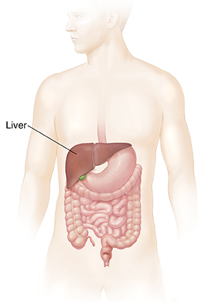
Understanding Radiofrequency Ablation (RFA) of Liver Tumors
Radiofrequency ablation (RFA) is a way to treat small liver tumors. RFA uses heat to kill the tumor cells. A high-frequency electrical current is sent into the tumor through a needle-like probe that holds the electrodes. This creates heat that kills the tumor cells without harming a lot of nearby healthy cells.

How to say it
RAY-dee-oh-FREE-kwehn-see uh-BLAY-shuhn
Why RFA of liver tumors is done
Radiofrequency ablation may be done if you can't have liver surgery. Or it may be done along with surgery.
It’s most often done if you have a small number of tumors in your liver and the tumors are small (up to about 3 centimeters across). The tumors can't be near bile ducts, major blood vessels, the diaphragm (the muscle that helps you breathe), or other nearby organs.
How RFA of liver tumors is done
This treatment is done in a hospital, and takes 1 to 3 hours. During the procedure:
-
A small tube (IV) will be put into a vein in your hand or arm. It's used to give you medicine (anesthesia) to make you relax or sleep through the treatment. A thin needle might be used to put numbing medicine into the skin where cuts will be made.
-
Grounding pads will be stuck to another place on your skin to help control the electric current that's used in RFA.
-
The healthcare provider makes a small cut (incision) in your skin. In some cases, the provider makes a few cuts and a long, thin tube with a tiny light and camera on the end (laparoscope) is put through 1 of the cuts. This helps the provider see inside your body. In other cases, the provider may make 1 large cut instead.
-
The provider puts a needle-like probe through the cut and into your liver. The probe may have 1 or many electrodes.
-
An imaging scan, like ultrasound, MRI, or CT, is used to guide the probe into the tumor.
-
When it's in the right place, electricity is sent through the electrode. This creates heat that kills the tumor cells. This is done for each tumor.
-
The tools are then taken out, and the cuts in the skin are bandaged. They seldom need stitches (sutures). The grounding pads are taken off.
-
You'll wake up in a recovery room. You may be attached to machines that keep track of your blood pressure, breathing, oxygen level, and heart rate. If needed, you'll be given medicines for pain or nausea.
-
When you fully wake up after treatment, you may be able to go home. Or you might stay in the hospital for a night or so.
-
A CT or MRI will be done soon after RFA to see if there are any tumors left in your liver. This will be repeated every 3 to 4 months to watch for new tumors. RFA can be repeated, if needed.
Risks of RFA of liver tumors
Any medical procedure carries some risk, but seldom causes problems. Possible risks of RFA include:
-
Problems from the anesthesia
-
Pain (often shoulder pain)
-
Skin burns
-
Bleeding
-
Leaking of bile from the liver
-
Infection
-
Blood clots
-
Damage to nearby organs, such as the gallbladder, bile ducts, intestines, or diaphragm
-
Damage to nearby blood vessels
-
Liver abscess, which is a pocket of infection inside the liver
-
Aches, fever, chills, delayed pain, and upset stomach (nausea) for a few days (called postablation syndrome)
Online Medical Reviewer:
Jessica Gotwals RN BSN MPH
Online Medical Reviewer:
Rajadurai Samnishanth Researcher
Online Medical Reviewer:
Susan K. Dempsey-Walls RN
Date Last Reviewed:
12/1/2024
© 2000-2026 The StayWell Company, LLC. All rights reserved. This information is not intended as a substitute for professional medical care. Always follow your healthcare professional's instructions.